

On this page
Real microscopy, annotated structures and measurements
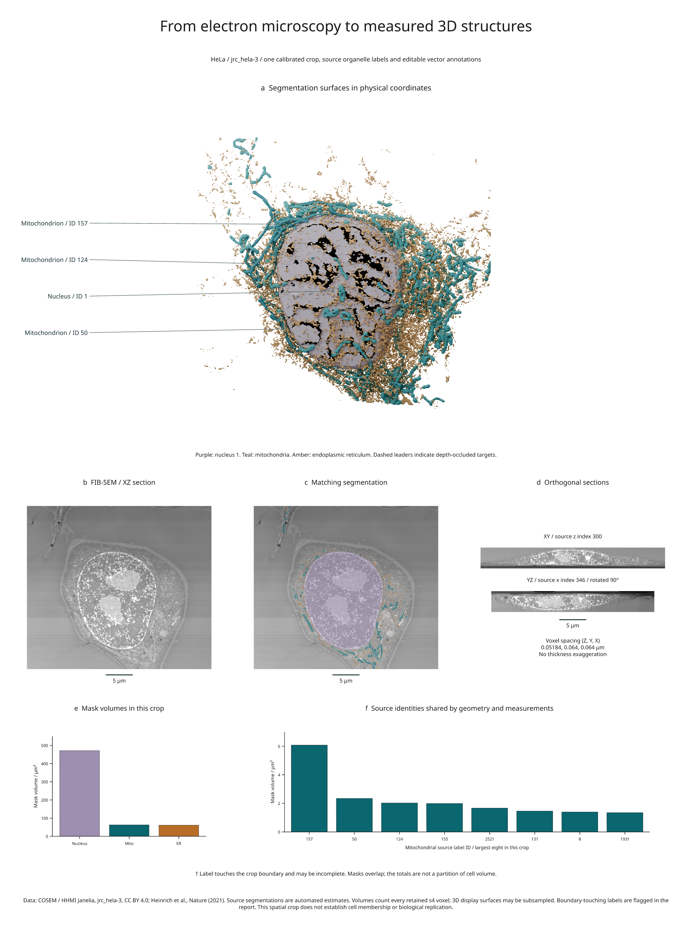
This figure combines FIB-SEM microscopy, source organelle segmentations, calibrated orthogonal sections and quantitative summaries from one fixed spatial crop of jrc_hela-3. The data are real; the labels are the source's automated estimates.
Full-size figure · Executable recipe · Measurements and render evidence · Source checksums

Reproduce it
From 4.3 the example imports from inklet.volume; the inklet.experimental
paths still work and, from 4.4, warn.
Use the v4.9.0 release checkout and Blender 4.2 or 4.5 LTS:
git clone --branch v4.9.0 https://github.com/Mapika/inklet.git
cd inklet
python -m pip install -e '.[volume,render]'
python -m pip install -r examples/biology/requirements.txt
python examples/real_biology.py --blender /path/to/blender
The recipe downloads approximately 56.6 MB of hash-locked source objects. It reads N5 through the separately pinned example dependencies, extracts meshes, builds a Blender scene and renders with an available GPU by default. CPU fallback occurs during device discovery. The core volume API itself performs no downloads.
Outputs in out/real-biology/ include the source cache, meshes, .blend scene,
SVG/PDF/PNG figure, review HTML and measurements.json. The source reader checks
that all four arrays have matching shapes and physical transforms. Every
available downloaded object must match its recorded size and SHA-256. Absent
N5 background chunks are recorded explicitly. Meshes and the scene recipe are
fingerprinted so a data or geometry change rebuilds the scene before rendering.
Coordinates and provenance
The source level is s4, shape 750 × 62 × 775 in ZYX order. Spacing is
51.84 × 64 × 64 nm, with a centre-origin translation of 24.3 × 30 × 30 nm
in ZYX order. The recipe converts these coordinates to micrometres without
stretching any dimension.
The half-open crop is Z[40:640], Y[0:62], X[80:630]. Its physical extent is
31.104 × 3.968 × 35.2 µm in ZYX order. The original world origin is retained.
The field includes source labels around the central nucleus; the crop does not
infer which cell owns each organelle or establish complete cell coverage.
- Panel a renders source segmentation surfaces. Callouts use original IDs and actual mesh surface points. Occluded anchors use dashed leaders.
- Panels b and c show the same XZ section at source Y index 31. They share the
explicit intensity window
[17000, 26000]. The overlay uses 55% mask color. - Panel d shows physical XY and YZ sections. YZ is rotated 90° for layout; its geometry is not stretched. Scale bars show 5 µm.
- Panels e and f count every retained source-level voxel. They use the same segmentation arrays as the displayed geometry.
Recorded measurements
| Mask | Foreground voxels in crop | Voxel-count volume |
|---|---|---|
| Nucleus, ID 1 | 2,218,929 | 471.1599 µm³ |
| Mitochondria, all retained IDs | 296,336 | 62.9230 µm³ |
| Endoplasmic reticulum, all retained IDs | 289,154 | 61.3980 µm³ |
The crop retains 209 mitochondrial label IDs and 58 ER label IDs. These are source labels, not verified counts of independent biological organelles. Of the eight largest retained mitochondrial labels, 252 and 193 touch the crop boundary; their bars are marked with a dagger. Their volumes may be incomplete.
The masks are not disjoint: the nucleus/ER pair overlaps at 80,697 voxels, and the nucleus/mitochondria and mitochondria/ER pairs overlap at 14 and 13 voxels. The recipe preserves and reports these overlaps instead of modifying the source to make a partition. Do not sum the bars into a cell volume.
What the rendering establishes
The oblique-section example extends this dataset with two angled physical planes shared by 3D annotations, image panels, label-area comparisons and marked intensity profiles.
The example checks that source IDs, physical coordinates, slices and reported
values remain connected through the figure pipeline. It is not a reanalysis
of the paper's biological findings or a validation of its segmentation models.
The source s4 level is already coarse relative to acquisition resolution.
The three individually annotated mitochondrial meshes use every s4 sample.
The nucleus, remaining mitochondria and ER use display stride 2, which can lose
thin structures. Measurements still use every retained voxel. Boundary-touching
aggregate surfaces explicitly permit artificial caps. The report records mesh
sizes, stride, cap status and the actual Blender device and camera-plan evidence.
Attribution and reuse
Data: COSEM Project Team / HHMI Janelia Research Campus, accessed 7 September 2026 through the COSEM data registry, which declares CC BY 4.0. Publication: Heinrich et al., Whole-cell organelle segmentation in volume electron microscopy, Nature 599, 141–146 (2021).
The figure changes the presentation through cropping, windowing, overlays, surface extraction, rendering, labels and quantitative summaries. The derived figure retains CC BY 4.0; the example code is MIT. Raw data and generated meshes are downloaded locally and are not bundled with Inklet. See third-party notices.